- Radiological imaging
There are various radiological procedures that may be done when an individual is diagnosed to have chronic kidney disease. These tests are done when the doctor suspects that you may have certain diseases that can be detected by the procedure, for example, stone disease, tumor, structural abnormalities, etc. However, the tests may sometimes be ordered to gain more information about the size and condition of the kidneys.
What is an ultrasound of the kidney?
During an ultrasound examination, a small device known as a probe sends sound waves into the area of interest, and images are recorded on a computer. The black-and-white images show details of internal structures such as muscles, organs, and blood vessels.
A simple ultrasound of the kidneys can provide information such as the size of the kidneys whether there is obstruction from stones or tumors or abnormal structures like cysts or scarring.
This test is pain-free although some pressure can be felt at times. It is also free of radiation. The procedure usually takes about 30 minutes.
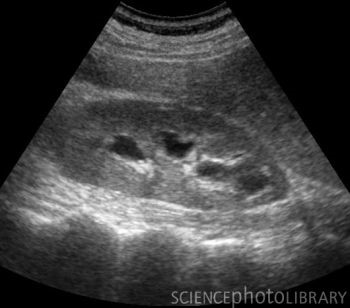
Diagram 1: Image of an enlarged kidney on ultrasound
How to prepare for an ultrasound examination?
An ultrasound examination is usually done in the radiology department, but it may also occasionally be performed at the bedside in wards.
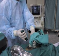
You need to have a full bladder so you must not pass urine just before the examination. You will be asked to change into a cloth gown and lie on a table. The room is usually dark so the images can be seen clearly on the computer screen. A technician (sonographer) or a doctor will spread a clear, warm gel over the lower ribs at the back and in front of the abdomen. This gel helps with the transmission of the sound waves.

Diagram 2: Illustration of an ultrasound examination
What are the other types of radiology imaging that may be required?
Other tests may include:
- Computed tomography scan (CT scan)
- Magnetic Resonance Imaging scan (MRI)
- Intravenous Pyelogram/Urogram
Some of this radiological imaging may involve exposure to radiation and /or require the use of contrast agents. Contrast agents can cause further deterioration of kidney function in certain situations such as individuals with impaired kidney function, dehydration or advanced age > 75 years. In such situations, special precautions will be required
Please alert the doctor should you
- Be pregnant.
- Have any history of
- Discussion with your doctor is important. In some cases, an alternative investigation may be chosen, or the risk of an allergic reaction can be reduced with as a short-course of steroids before exposure to the contrast agents
- Kidney biopsy
In some cases, the doctor may want to examine a small sample of your kidney under a microscope to determine the underlying cause and extent of disease.
How is a kidney biopsy done?
The procedure of getting a tiny sample of tissue from the kidney is called a biopsy. This is a minor procedure which is done under aseptic technique. A local anaesthetic agent is used to provide pain relief.

The procedure is carried out with the patient lying on his/ her abdomen under ultrasound guidance (used to locate the kidney). Occasionally, a biopsy is done under CT scan guidance.
A local anaesthetic agent is injected into the area for biopsy to provide pain relief. A special needle is used to obtain the tiny tissue sample.
After the kidney biopsy, your blood pressure, pulse rate and pain score are monitored at regular intervals. You will also be monitored for signs of bleeding.
What are the possible complications of a renal biopsy?
- Pain at the site of the biopsy.
- Bleeding – the most common complication. However, it is most often minor
- Passing blood in the urine – usually transient
- Haematoma – a collection of blood around the kidney. Commonly small and do not cause symptoms.
What are the contraindications for a renal biopsy?
- Bleeding problems
- Uncontrolled blood pressure
- Uncooperative patient
- Urinary tract infections or skin infections over the intended biopsy site
- Others : Usually relative contraindications
- A single functioning kidney
- Certain anatomical abnormalities of the kidney
- Pregnancy
- Obesity
When would you need a kidney biopsy?
When you have
- Haematuria (blood in the urine) in those with progressive kidney disease i.e. increasing proteinuria, worsening blood pressure or loss of kidney function
- Proteinuria ( protein in the urine) – in those with relatively high or increasing levels of protein in the urine, and in those with progressive kidney disease
- Unexplained kidney failure
Apart from providing information for the diagnosis of kidney disease, another reason for performing a kidney biopsy may be to monitor the amount of damage in the kidneys, and assess the effect of treatment that has been given.
Source:
Diagram 1: http://www.sciencephoto.com/image/98454/350wm/C0034228-Enlarged_kidney,_ultrasound_scan-SPL.jpg
Diagram 2: http://www.childrenscolorado.org/imgs/KidsHealth/image/ial/images/871/871_image.gif
| Last Reviewed | : | 3 May 2016 |
| Writer | : | Dr. Anita Bhajan Manocha |
| Accreditor | : | Dr. Sunita Bavanandan |

